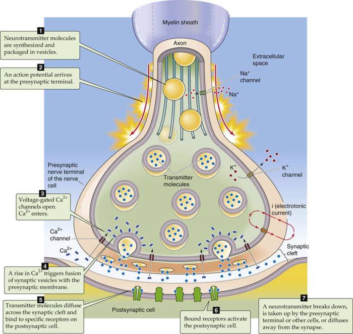
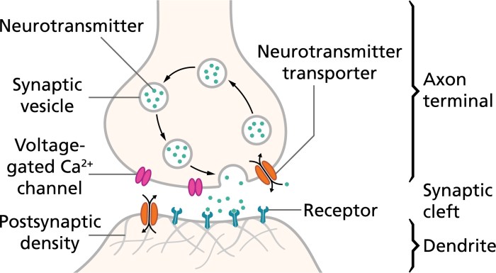
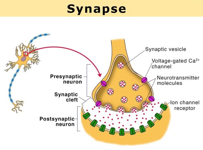
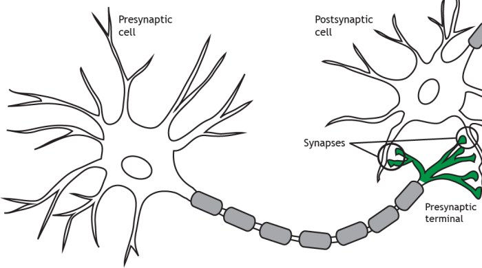
Figure 7-2 is a diagram of a synapse, the fundamental unit of communication between neurons. It provides a comprehensive visual representation of the synapse’s structure, function, and significance in neurotransmission. By examining this diagram, we embark on a journey into the intricate world of synaptic signaling, uncovering the mechanisms that govern neuronal communication and the complexities of the human brain.
The diagram showcases the pre-synaptic terminal, post-synaptic terminal, and synaptic cleft, highlighting their distinct roles in neurotransmitter release, binding, and signal transduction. It further illustrates the process of synaptic plasticity, a dynamic phenomenon that underlies learning and memory. Through a detailed analysis of Figure 7-2, we gain a profound understanding of synaptic function and its implications for neurological disorders and therapeutic interventions.
Diagram Overview
Figure 7-2 presents a comprehensive diagram of a synapse, providing insights into the structure and function of this critical neural junction. The diagram illustrates the key components of a synapse, including the pre-synaptic terminal, post-synaptic terminal, and synaptic cleft, and their arrangement within the synapse.
Synapse Structure

Pre-Synaptic Terminal
The pre-synaptic terminal contains synaptic vesicles filled with neurotransmitters. When an action potential arrives at the pre-synaptic terminal, it triggers the release of neurotransmitters into the synaptic cleft.
Post-Synaptic Terminal
The post-synaptic terminal contains receptors that bind to neurotransmitters released from the pre-synaptic terminal. Binding of neurotransmitters to receptors opens ion channels, allowing ions to flow into or out of the post-synaptic neuron, generating a post-synaptic potential.
Synaptic Cleft
The synaptic cleft is the space between the pre-synaptic and post-synaptic terminals. It plays a crucial role in neurotransmission by allowing neurotransmitters to diffuse across the gap and bind to receptors on the post-synaptic terminal.
Neurotransmitter Release and Binding

Neurotransmitter Release
Neurotransmitter release from the pre-synaptic terminal is a calcium-dependent process. When an action potential arrives at the pre-synaptic terminal, it causes an influx of calcium ions, which triggers the fusion of synaptic vesicles with the pre-synaptic membrane and the release of neurotransmitters into the synaptic cleft.
Neurotransmitter Binding
Neurotransmitters released from the pre-synaptic terminal diffuse across the synaptic cleft and bind to receptors on the post-synaptic terminal. The binding of neurotransmitters to receptors opens ion channels, allowing ions to flow into or out of the post-synaptic neuron, generating a post-synaptic potential.
Synaptic Plasticity

Definition
Synaptic plasticity refers to the ability of synapses to change their strength over time in response to experience. Synaptic plasticity is a fundamental mechanism underlying learning and memory.
Forms of Synaptic Plasticity
There are two main forms of synaptic plasticity: long-term potentiation (LTP) and long-term depression (LTD). LTP is a strengthening of the synapse, while LTD is a weakening of the synapse.
Role in Neural Circuit Development and Function
Synaptic plasticity plays a crucial role in neural circuit development and function. It allows the brain to adapt to new experiences and to store memories.
Comparison with Other Synapse Diagrams
Other Common Synapse Diagrams
There are several other commonly used diagrams of synapses, including the “ball-and-stick” model and the “ribbon” model.
Comparison with Figure 7-2
Figure 7-2 is a simplified diagram of a synapse that clearly illustrates the main components and their arrangement. It is well-suited for teaching and general understanding of synapse structure and function.
Applications in Neuroscience: Figure 7-2 Is A Diagram Of A Synapse

Understanding Synaptic Function and Dysfunction
Figure 7-2 can be used to understand synaptic function and dysfunction in various neurological disorders, such as Alzheimer’s disease and Parkinson’s disease.
Developing Therapeutic Interventions, Figure 7-2 is a diagram of a synapse
Figure 7-2 can inform the development of therapeutic interventions for synapse-related disorders. For example, drugs that enhance LTP could be used to treat memory deficits in Alzheimer’s disease.
Advancements in Neuroscience Research
Figure 7-2 has contributed to advancements in neuroscience research by providing a clear and concise representation of synapse structure and function. It has been used in numerous studies to investigate the role of synapses in learning, memory, and disease.
Question Bank
What is the significance of Figure 7-2?
Figure 7-2 is a comprehensive diagram that provides a detailed visual representation of the synapse’s structure, function, and role in neurotransmission. It serves as a valuable educational tool and a reference for neuroscientists and researchers.
How does Figure 7-2 illustrate synaptic plasticity?
The diagram depicts the dynamic nature of synaptic plasticity by highlighting the changes that occur in synaptic strength over time. It illustrates the processes of long-term potentiation and long-term depression, which are crucial for learning and memory.
What are the applications of Figure 7-2 in neuroscience?
Figure 7-2 is widely used in neuroscience research to understand synaptic function and dysfunction in various neurological disorders. It aids in the development of therapeutic interventions for synapse-related disorders and contributes to advancements in our understanding of brain function.